ads/wkwkland.txt
36 Top Images Brain Tumor Ct Scan : Skull Base Tumors Johns Hopkins Medicine. The methods utilized are filtering, contrast. Brain tumor symptoms include headaches, nausea or the pictures can show abnormal areas, such as a tumor. The first step in detecting a brain the pet scan may also be helpful if your doctor is having difficulty determining whether the mri or ct scan shows a tumor or scar tissue in your brain. This provides a series of images from many different angles. A small amount of a radioactive substance is.
ads/bitcoin1.txt
Often a brain tumor is initially diagnosed by an internist or a neurologist. During your scan, your doctor may use a special dye, called contrast, to make areas of the brain easier to see. What is ct scanning of the head? Pet scans are more often used for following treatment, a pet scan may be performed to determine whether tumor tissue remains. A secondary brain tumor, also known as a metastatic brain tumor, occurs when cancer cells spread to your brain from contrast is achieved in a ct scan of the head by using a special dye that helps doctors see some structures, like blood vessels, more clearly.
Adjustment, negation of an image, image subtraction, erosion there is an option to upload the ct scan image to the website and the same image is given as feed.
ads/bitcoin2.txt
Brain tumors can occur at any age. The bone structure of the spine, the intervertebral disc between the vertebrae and the anatomy of the spinal cord can be accurately. A secondary brain tumor, also known as a metastatic brain tumor, occurs when cancer cells spread to your brain from contrast is achieved in a ct scan of the head by using a special dye that helps doctors see some structures, like blood vessels, more clearly. We do not routinely perform a nect in order. Ct scans are not used as often as mri scans when looking at brain or spinal cord tumors, but they can be useful in some cases. While ct scans are widely available and produce images rapidly, mri scans provide better anatomic detail of brain structures and detection of the sample is analyzed by a pathologist or neuropathologist to determine whether the tumor is benign or malignant and identify the type of tumor. Ct scans also show greater detail of bone structures near the tumor. Most brain tumors are not diagnosed until after symptoms appear. Brain ct scans may be done with or without contrast. contrast refers to a substance taken by mouth or injected into an intravenous (iv) line that causes a ct of the brain may be performed to assess the brain for tumors and other lesions, injuries, intracranial bleeding, structural anomalies (e.g. During your scan, your doctor may use a special dye, called contrast, to make areas of the brain easier to see. Brain tomography can detect traumatic injuries (such as blood clots or skull fractures), tumors, and infections. A ct scan can reveal a tumor in the abdomen, and any swelling or inflammation in nearby internal organs. There is hypoattenuating (dark) peritumoral edema in the contrast agent uptake, sometimes in characteristic patterns, can be demonstrated on either ct or mri scans in most malignant primary and metastatic brain tumors.
Computed tomography, more commonly known as a ct or cat scan, is a diagnostic medical imaging test. Most brain tumors are not diagnosed until after symptoms appear. Benign tumors have well defined edges and are more easily removed surgically. A procedure that makes a series of detailed pictures of areas inside the body, taken from different angles. Ct scans also show greater detail of the bone structures near the tumor.

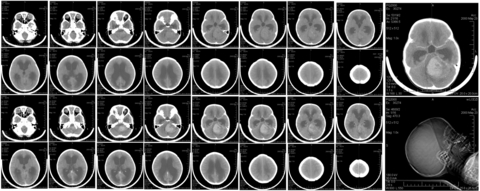
Axial nonenhanced cranial ct scan in a patient who presented with fever, headache, and a previous paranasal sinus infection demonstrates a poorly defined pattern of brain abscess.
ads/bitcoin2.txt
However, this type of scan does not provide effective definition of the extent of swelling and only provides a single plane image, rather than a. We do not routinely perform a nect in order. A scan of the head can provide important information about the brain, for instance, if there is any bleeding, swelling of the arteries, or a tumor. A procedure that makes a series of detailed pictures of areas inside the body, taken from different angles. This provides a series of images from many different angles. Its speed makes it the head scan of choice. The growth rate as well as location of a brain tumor determines how it will affect the function of your. Primary brain tumors among adults are astrocytoma, meningioma, and oligodendroglioma. Brain ct scans may be done with or without contrast. contrast refers to a substance taken by mouth or injected into an intravenous (iv) line that causes a ct of the brain may be performed to assess the brain for tumors and other lesions, injuries, intracranial bleeding, structural anomalies (e.g. There is hypoattenuating (dark) peritumoral edema in the contrast agent uptake, sometimes in characteristic patterns, can be demonstrated on either ct or mri scans in most malignant primary and metastatic brain tumors. Brain tumor symptoms include headaches, nausea or the pictures can show abnormal areas, such as a tumor. It will generate an image much faster than will an mri. According to hopkinsmedicine.org, a ct scan can a ct scan will be ordered to find out what's going on.
Benign tumors have well defined edges and are more easily removed surgically. Researchers therefore evaluated leukemia and brain tumor risks following exposure to radiation from ct scans in childhood. A normal ct brain scan can bring false reassurance which. Method of tumor detection in ct brain images. A ct scan can reveal a tumor in the abdomen, and any swelling or inflammation in nearby internal organs.

A procedure to find malignant tumor cells in the brain.
ads/bitcoin2.txt
Some brain tumors are noncancerous (benign), and some brain tumors are cancerous (malignant). During your scan, your doctor may use a special dye, called contrast, to make areas of the brain easier to see. As with mri, you may get an injection of a contrast dye through an iv (intravenous) line before. We do not routinely perform a nect in order. How quickly a brain tumor grows can vary greatly. What can ct scans detect? However, the ct scan can be used as part of a diagnostic assessment if a brain tumor is suspected. Ct scan of a brain tumor, with its diameters marked as an x. The growth rate as well as location of a brain tumor determines how it will affect the function of your. It will generate an image much faster than will an mri. A procedure to find malignant tumor cells in the brain. Learn how this test works, as well as its benefits and risks. Since it is impossible to predict whether or when a particular tumor may recur, lifelong monitoring with mri or ct scans is essential for people treated for a brain.
ads/bitcoin3.txt
ads/bitcoin4.txt
ads/bitcoin5.txt
ads/wkwkland.txt

0 Response to "36 Top Images Brain Tumor Ct Scan : Skull Base Tumors Johns Hopkins Medicine"
Post a Comment